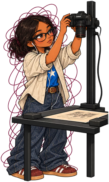
From specimen to record.

Preparing, imaging and rehousing dragonfly specimens for the DIODE digitization project at the American Museum of Natural History.

Role
Collections Digitization Volunteer
Period
2026–present
Lab
Jessica Ware’s lab · Invertebrate Zoology, AMNH
Supervisors
Ellie Gamett and Rin Krichilsky

Bringing the collection into view

A specimen holds more than its appearance. Its collection labels carry context that needs to remain connected to it as the collection is digitized.

I volunteer for a few hours each week, working through the physical preparation, image capture, review and rehousing involved in that process. The setup combines careful handling with 3D-printed trays, a motorized imaging stage and BugBase, the existing software used to coordinate capture.

  1. CameraPhotographs specimens and their associated labels.
  2. LightingIlluminates the imaging area.
  3. Motorized stagePositions the loaded tray beneath the camera.
  4. Prepared trayKeeps specimens and associated labels organized for capture.One tray position, enlarged
  5. Control softwareBugBase coordinates the imaging sequence.Its two screens, capture and review
  6. Waiting specimensPrepared batches ready for imaging.
The imaging workstation. An illustrated view of the existing digitization setup.
  1. 01 · Preparing the specimens

    I retrieve specimens and their associated labels, organize them in the trays, and add unique identifiers. Some specimens have detached parts, which I carefully position for imaging following the lab’s handling procedure. I also brush loose debris from the tray before capture.
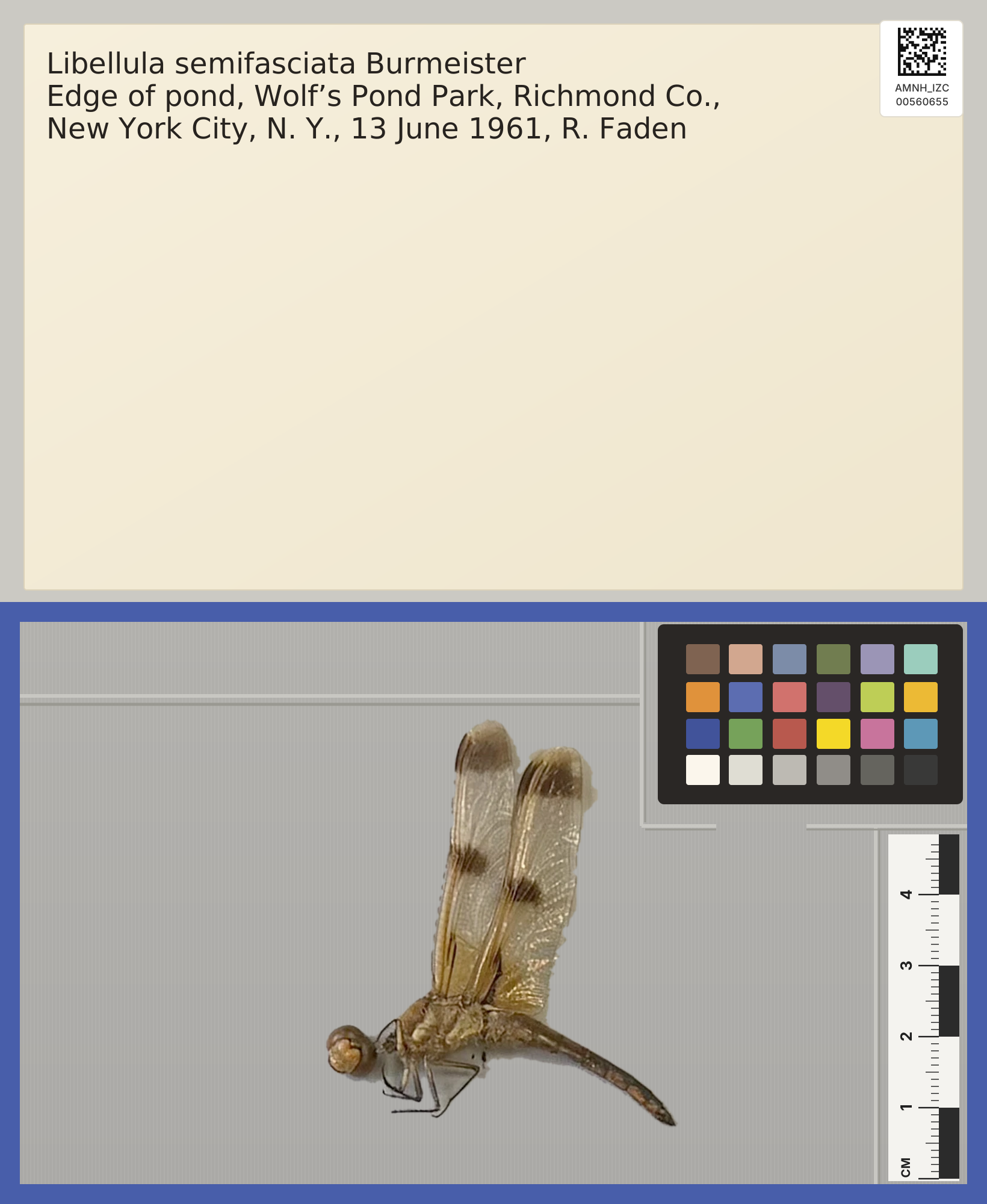
    1. Specimen and labelThe specimen and its collection label remain associated throughout the process.
    2. IdentifierA unique identifier connects the specimen with its digital images and records.
    3. Color targetThe color target provides a reference for interpreting image color.
    4. ScaleThe scale provides a size reference in the captured image.

    Keeping the specimen, label and identifier associated is part of the preparation—not something to sort out after the photographs are taken.
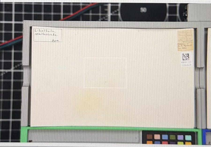
    One tray position. An illustrated reconstruction, redrawn from a photograph: the label is transcribed, the identifier regenerated from its scanned value, and the specimen is the real photograph. Not an original digitization output.
  2. 02 · Running and reviewing the capture

    Once the tray is ready, I load it onto the imaging stage and use BugBase to run the capture sequence. The stage positions the tray beneath the camera so specimens and their labels can be photographed.

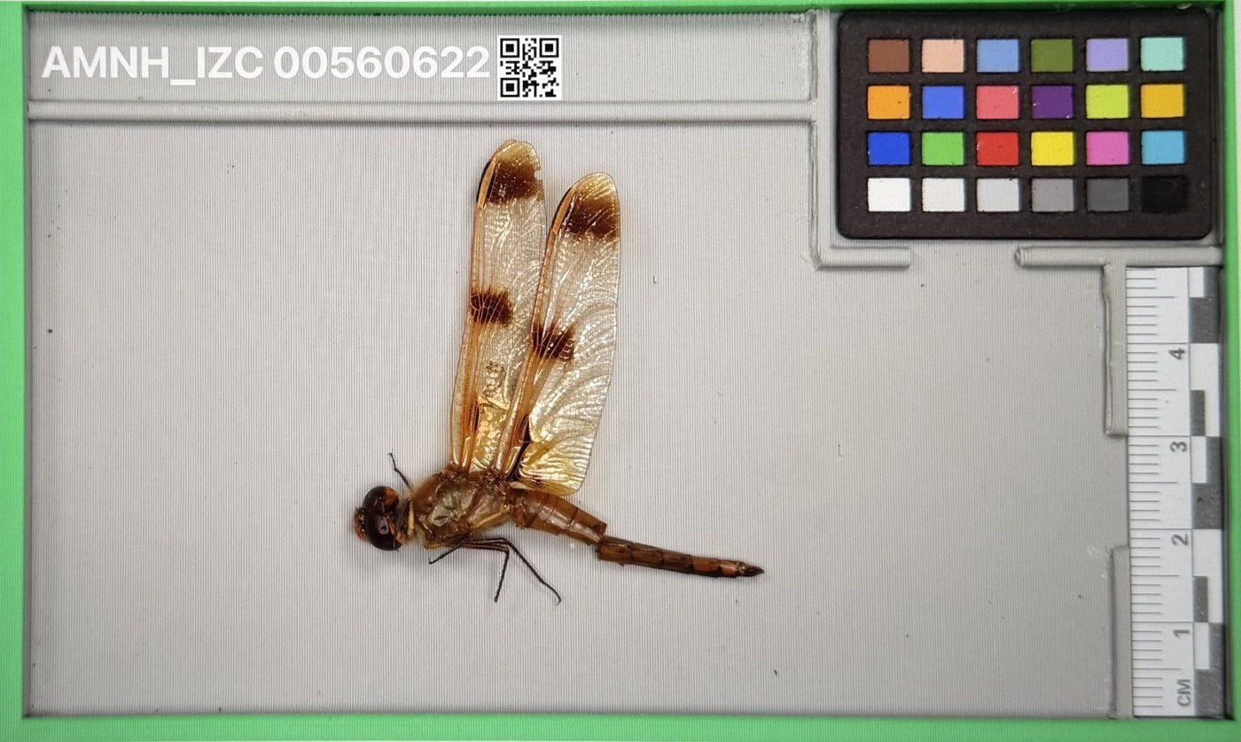
    I then review the captured specimen-and-label pairs, checking the images and approving or rejecting them before continuing. The capture sequence is automated in part; preparation and image review still depend on attention to each specimen.

    BugBase on the workstation’s laptop. Two of its screens, recreated as illustrations.
    Camera control
    I use BugBase to run the imaging sequence and monitor the camera view.An illustrative recreation of the CameraControl window, simplified from a photograph of the screen.
    Image review
    I check the captured specimen-and-label pairs and approve or reject images before continuing.An example saved specimen image, with its identifier, scale and color reference visible (AMNH_IZC 00560622, a different specimen from the tray position above). The window is an illustrative recreation; the label image isn’t shown.
  3. 03 · Rehousing after imaging

    After imaging, I rehouse the specimens in new sleeves with their associated collection material. The work ends with both a digital image record and the physical specimen ready to return to the collection.

What draws me to it

Working through the process has made me more attentive to what sits behind a digital collection: careful handling, readable labels, consistent images, and decisions made one specimen at a time.

I’ve also been exploring the existing BugBase code to understand how the software coordinates the workflow. I’m interested in the tools, but equally in learning about the collection and the research those records can support.

Lab and tools

This work takes place in Dr. Jessica L. Ware’s lab in AMNH’s Division of Invertebrate Zoology, under the supervision of Ellie Gamett and Rin Krichilsky, doctoral researchers affiliated with Columbia University and AMNH.

I use existing tools and procedures developed by the project team and collaborators, including BugBase, the imaging rig and the 3D-printed trays. My contribution is preparing specimens, operating the imaging workflow, reviewing images and rehousing collection material.